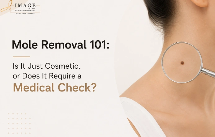
Most people who book a mole removal appointment in Kolkata arrive with one of two concerns. Either they have a mole they find aesthetically bothersome and want removed. Or they have noticed a change in a mole — in its size, shape, colour, or surface texture — and they are not sure whether to have it assessed by a dermatologist or removed without further investigation. The answer to both questions begins with the same fundamental step: a clinical assessment. Not because every mole is dangerous, but because no one — not the patient, and not a non-clinical practitioner — can confidently distinguish a benign mole from an early melanoma by visual inspection alone.
This guide from IMAGE Clinic explains the difference between cosmetic mole removal and medically indicated mole assessment, how dermatologists determine which approach is appropriate, and what to look for in a mole that warrants urgent clinical attention.
| Considering mole removal? The first step is always a clinical assessment. Book yours at IMAGE Clinic. Book Your Mole Assessment at IMAGE Clinic |
IMAGE Clinic, Kolkata — Mole Assessment and Removal
IMAGE Clinic is a dermatologist-led skin clinic in Kolkata performing mole assessment and mole removal with dermoscopy at every consultation. Board-certified dermatologists assess every lesion clinically before recommending any removal technique. View our team and documented patient results for reference.
The Essential First Question: Is This Mole Safe to Remove?
Before any discussion of technique, timing, or cost, a single clinical question must be answered: is this mole benign? If the answer is a confident clinical yes — confirmed by dermoscopy — the conversation about cosmetic removal can proceed. If there is any clinical uncertainty, the appropriate course of action is excision biopsy with histopathological analysis, not cosmetic removal.
This distinction matters enormously because cosmetic laser or radiofrequency ablation destroys the mole tissue. If that tissue contains malignant cells, the destruction of the lesion eliminates the tissue needed for a diagnosis that could determine the patient’s cancer staging and treatment. A cosmetically removed melanoma is a missed opportunity for early intervention that has potentially serious consequences.
No reputable dermatologist will agree to remove a mole — by any method — without first performing a clinical assessment that confirms the lesion is benign and appropriate for cosmetic removal.
How Dermatologists Assess a Mole: Dermoscopy and the ABCDE Criteria
The ABCDE Clinical Criteria
The ABCDE criteria are the internationally established clinical framework for evaluating whether a pigmented skin lesion requires further investigation:
- A — Asymmetry: A benign mole is typically symmetrical — if you drew a line through the centre, both halves would match. Asymmetry in either axis is a clinical flag.
- B — Border: Benign moles have clearly defined, regular borders. Irregular, scalloped, or poorly defined borders are concerning.
- C — Colour: A benign mole is uniform in colour — typically a consistent brown or tan. Multiple colours within the same mole (varying shades of brown, black, red, white, or blue) are a warning sign.
- D — Diameter: Moles greater than 6mm in diameter — approximately the size of a pencil eraser — are more likely to require further assessment, though smaller lesions can also be concerning if other criteria are met.
- E — Evolution: Any change in a mole over time — in size, shape, colour, surface texture, or new symptoms such as itching, bleeding, or crusting — is the most clinically significant flag and should prompt immediate dermatological assessment.
Dermoscopy: What Visual Inspection Cannot Show
Dermoscopy is a non-invasive magnification technique that allows dermatologists to examine the structural features of a pigmented lesion at 10–20x magnification under polarised light. Dermoscopy reveals the internal architecture of the mole — the pigment network, vascular pattern, the distribution of colour, and specific structures associated with malignancy — that are not visible to the naked eye.
At IMAGE Clinic, dermoscopy is performed at every mole assessment. It significantly improves the accuracy of clinical diagnosis — allowing the dermatologist to confidently classify a lesion as benign or to identify features that require histopathological analysis. Mole removal without dermoscopy is a lower standard of care, and IMAGE Clinic does not perform mole removal in Kolkata without this assessment step.
When Mole Removal Is Cosmetic: What the Process Involves
For moles confirmed as benign by dermoscopy and clinical assessment, the appropriate removal technique is selected based on the mole’s type, depth, size, and location. The most common techniques at IMAGE Clinic are:
Laser Ablation (CO2 or Er: YAG)
Laser ablation vaporises the mole tissue layer by layer with precision. For flat or superficial moles confirmed as benign junctional nevi or seborrhoeic keratoses, this is the technique of choice — producing the most cosmetically acceptable result with minimal scar risk. The procedure takes ten to twenty minutes under topical anaesthetic. Healing involves a small crusting area over seven to fourteen days. Laser skin resurfacing technology is also employed for flat pigmented lesions across the skin surface.
Radiofrequency (RF) Ablation
For raised, dome-shaped, or pedunculated moles — particularly intradermal nevi — RF ablation uses high-frequency electrical current to cut and coagulate the mole tissue with precise control. RF ablation is particularly useful for moles in cosmetically sensitive areas such as the face and neck, where minimal scarring is the priority.
Surgical Excision
For moles that are deeper, larger, or where dermoscopy identifies any feature warranting histopathological analysis, surgical excision under local anaesthetic is performed with the tissue submitted for pathological examination. This is the standard of care for any mole with clinical uncertainty — it enables both removal and diagnosis simultaneously. Patients who present at IMAGE Clinic with warts, skin tags, or other skin lesions alongside moles receive a comprehensive assessment of all lesions at the same appointment.
| Not sure whether your mole needs a medical check or cosmetic removal? Our dermatologists will assess it accurately. Book Your Mole Assessment |
What Happens After Mole Removal: Healing and Results
Post-removal care at IMAGE Clinic follows a standard protocol: keep the wound site clean and dry; apply the prescribed healing ointment; avoid sun exposure to the healing area for a minimum of four to six weeks; and attend the follow-up appointment at four to six weeks to assess healing and address any concerns.
The most common post-removal concern is post-inflammatory hyperpigmentation (PIH) at the healing site — a darker mark where the mole was, particularly in Indian skin. This is not the mole returning — it is a melanin response to the healing process. It can be treated with topical depigmenting agents and typically resolves within two to four months. Continued sun protection for at least three months post-removal is the most important factor in achieving the most cosmetically acceptable final result.
People Also Ask: Mole Removal in Kolkata
Is mole removal just cosmetic, or does it require a medical check?
Short answer: Every mole removal in Kolkata should begin with a medical assessment — regardless of whether the reason for removal is cosmetic or clinical concern. Dermoscopy is required to confirm a mole is benign before any cosmetic removal technique is applied. A mole removed cosmetically without assessment that turns out to be malignant represents a missed diagnostic opportunity with serious potential consequences.
When does a mole need to be checked by a dermatologist?
Short answer: Any mole that shows change — in size, shape, colour, surface texture, or sensation (itching, bleeding, crusting) — should be assessed urgently by a dermatologist. New moles appearing in adults over 40, moles larger than 6mm, moles with irregular borders or multiple colours, and any mole the patient has not seen before and cannot date should all be assessed before any removal is considered.
Is mole removal safe?
Short answer: Mole removal is safe when performed by a qualified dermatologist following clinical dermoscopic assessment. The primary safety requirement is confirming the lesion is benign before cosmetic removal. At IMAGE Clinic, all mole removal procedures are preceded by dermoscopy, and any mole with uncertain features is sent for histopathological analysis rather than cosmetically ablated.
What are the signs a mole is dangerous?
Short answer: The ABCDE criteria identify concerning moles: Asymmetry (uneven shape), Border irregularity, Colour variation within the mole, Diameter greater than 6mm, and Evolution (any change over time). The E criterion — change — is the most clinically significant. Any mole that has changed should be assessed by a dermatologist with dermoscopy before any other decision is made.
Does mole removal leave a scar?
Short answer: Scarring risk depends on the removal technique, mole depth, and the patient’s skin healing tendencies. Superficial laser ablation of benign flat moles typically heals with minimal to no visible scar. Surgical excision leaves a fine linear scar that fades progressively. The most common visible change after mole removal in Indian skin is post-inflammatory hyperpigmentation — which is treatable and temporary, not true scarring.
| Mole Removal in Kolkata — IMAGE Clinic Dermoscopy first. Clinical confidence. Safe, accurate results. Book Your Appointment Now | Call: 09830 836 666 Or visit your nearest IMAGE Clinic — find your nearest branch here. |